Internal Anatomy
As Elysia sp. is new to science, its internal anatomy has not yet been studied, but is expected to be similar to other species in the genus. For this reason, I have relied on detailed descriptions of the internal anatomy of other species within the genus to infer the likely internal anatomy of Elysia sp. It should be noted however that the internal anatomy, particularly reproductive anatomy, does vary throughout the genus, and so the information below should be carefully considered.
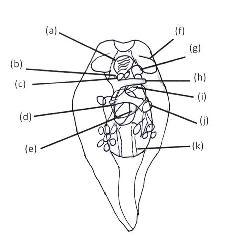
The diagram in Figure 1 below shows the internal anatomy a dissected E. australis, and it is clear from this diagram that the internal anatomy of this sea slug is relatively complex, particularly in the anterior end where a number of organs are condensed into quite a narrow area. It can be seen that the pharanyx (a), is located directly between the two rhinophores (f) of the animal and is quite ventral in placement. The eyes (b) are located posterior to the rhinophores, and though it is not clear in this diagram, they are on the uppermost dorsal surface. It should be noted that the position of the eyes in Elysia sp. differs from that of E. australis in that they are shifted ventrally to such a degree that they are more on the side of the animal than on the dorsal surface. The penis (g) is housed within a slit located posterior to the right rhinophore, as is common for all sacoglossans, whilst the vaginal opening (i) is situated just behind the intestine (h) and is closer to the mid line of the animal. The ampulla gland (j) is located posterior to and to the right of the vaginal opening. The prostate (k) is seen to be the most posterior organ, located behind, though not in close proximity to, the genital receptacle (e) which opens to the inner parapodial dorsal surface. The cerebral ganglion (c) is located immediately behind the pharanyx, approximately below the pericardium (not drawn on diagram) and although not entirely clear in this diagram, the mucus gland (d) is quite a large structure and located on the mid line of the animal between the cerebral ganglion and genital receptacle.

Figure 1- Internal anatomy of Elysia australis. Legend: a, pharanyx; b, eye; c, cerebral ganglion; d, mucus gland; e, genital receptacle; f, rhinophore; g, penis; h, intestine; i, vaginal opening; j, ampulla; k, prostate. Adapted from Jensen, 1992 [1].
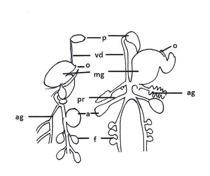
As stated above, the internal anatomy of the sea slugs can be quite complex and it is difficult to see many of the key reproductive structures in Figure 1. Figure 2 (below) provides a clearer illustration of the key reproductive anatomy within these sea slugs. It also demonstrates that despite their high anatomical complexity for such a relatively small animal, there is still a degree of variability in the layout of their internal anatomy. On the left is the reproductive anatomy for E. patina with E. timida on the right. It can be seen that much of the gross morphology is conserved between the two, but with some variations. The penes (p) of both species are located at the most anterior aspect, with the vas deferens (vd) leading down from the penis into the mucus gland (mg) in both. Both species also have the oviducal opening (o) to the right of the mucus gland, though it is shifted more toward the anterior aspect in E. timida. The albumen gland (ag) is located on the opposite side in these two species; on the left in E. patina and on the right below the mucus gland in E. timida. The ampulla (a) is seen to be opposite the albumen gland in both species, such that it is on the left in E. timida, and the right in E. patina. In E. timida the ampulla is preceded by the prostate gland; though not noted for E. patina, it would be reasonable to assume that a similar relationship would be seen in this species. Interestingly, the follicles (f) are seen to be slightly larger in E. patina, but more numerous in E. timida.

Figure 2- Reproductive anatomy varies throughout the Elysia. Schmatic diagrams of the reproductive anatomy of E. patina (left) and E. timida (right) demonstrate this variation. Legend: p, penis; vd, vas deferens; o, oviducal opening; mg, mucus gland; ag, albumen gland; pr, prostate; a, ampulla; f, follicle. E. timida diagram adapted from Jensen, 1992 [1], E. patina adapted from Jensen, 1986 [15].
|